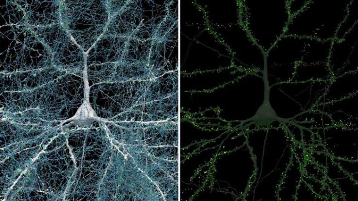
A team of scientists from Harvard and Google has created the most detailed 3D map of the human brain ever made. This map shows a very small piece of brain tissue, just one cubic millimeter in size, but it contains a huge amount of detail.
Within this tiny piece of the brain, there are about 57,000 cells, 230 millimeters of blood vessels, and almost 150 million synapses, which are the connections between brain cells.
To make this incredibly detailed map, the scientists had to cut the brain tissue into 5,000 very thin slices. They used a high-speed electron microscope to scan each slice.
After scanning, they used a machine-learning model to put all the slices back together electronically, like a puzzle, and to label all the different features. The final data set is enormous, taking up 1.4 petabytes of storage, which is a huge amount of data.
The focus of this map is on “connectomics,” which is the study of how cells in the brain are connected. This field is important because understanding these connections helps scientists learn how the brain processes information and stores memories. Creating this map was very complicated because the brain’s wiring is extremely intricate. However, the team’s work provides valuable insights into the brain’s structure and function.
The map is available for free on the Neuroglancer web platform, which means that researchers from all over the world can use it to study the human cortex in great detail. This opens up many opportunities for new discoveries in neuroscience, as scientists can explore the brain’s connections at a level of detail that was never possible before.


